Cardiac Organoids: Lifelike Functionality ▶ Experience the rhythmic pulseof our cardiac organoids, mimicking real human heart functionality for groundbreaking research.
Simplified Cultivation: Enhanced Efficiency ▶ Our cultivation process eliminates extracellular matrices, reducing variabilityand maximizing the efficiencyof functional cardiac organoid production.
Advanced Differentiation: Superior Maturation ▶ We've refined our differentiation methods to better replicate the human heart, achieving a precise balanceof myocardialand non-myocardial cells for more mature organoid functions.
Organism
Product Type
Organoid
Tissue
iPSC
Disease
Applications
Efficacy assay
Organoids are three-dimensional mini organs that mimic the functions of respective organs, reproducing their complex physiological structures and functions. Verify their efficacy with our advanced technology.
Toxicity test
Organoids are three-dimensional mini organs that mimic the functions of respective organs, reproducing their complex physiological structures and functions. Assess their toxicity with our advanced technology!
Innovate Cardiac Solutions with iPSC-Derived Organoids
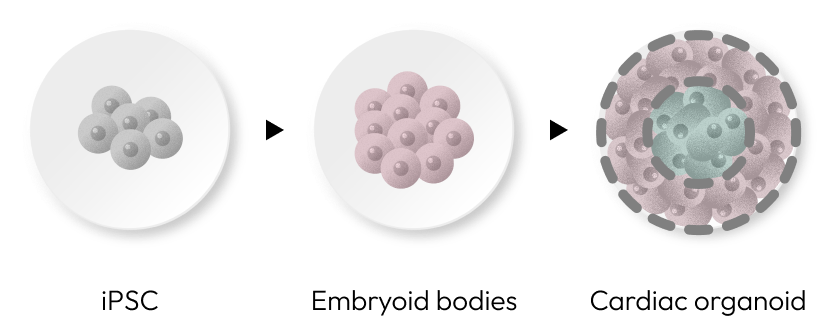
Leverage our breakthrough cardiac organoids, engineered from human-induced pluripotent stem cells (iPSCs). By transforming 3D embryoid bodies through precise signal manipulation, we create organoids that emulate heart tissue, advancing personalized cardiac research and therapy.
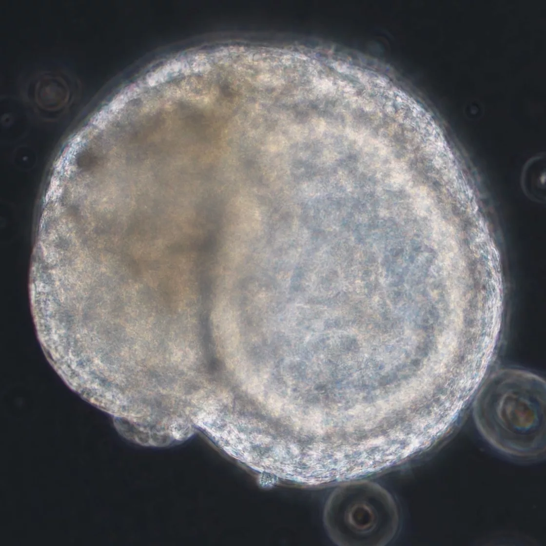
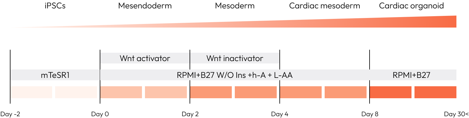
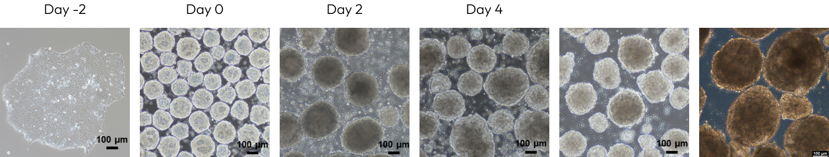
Generation of cardiac organoids
To create cardiac organoids, we utilized Wnt signaling activators and inhibitors, which are crucial for heart development, at various stages of differentiation within embryoid bodies. By day eight of differentiation, we observed spontaneous beating in the organoids.
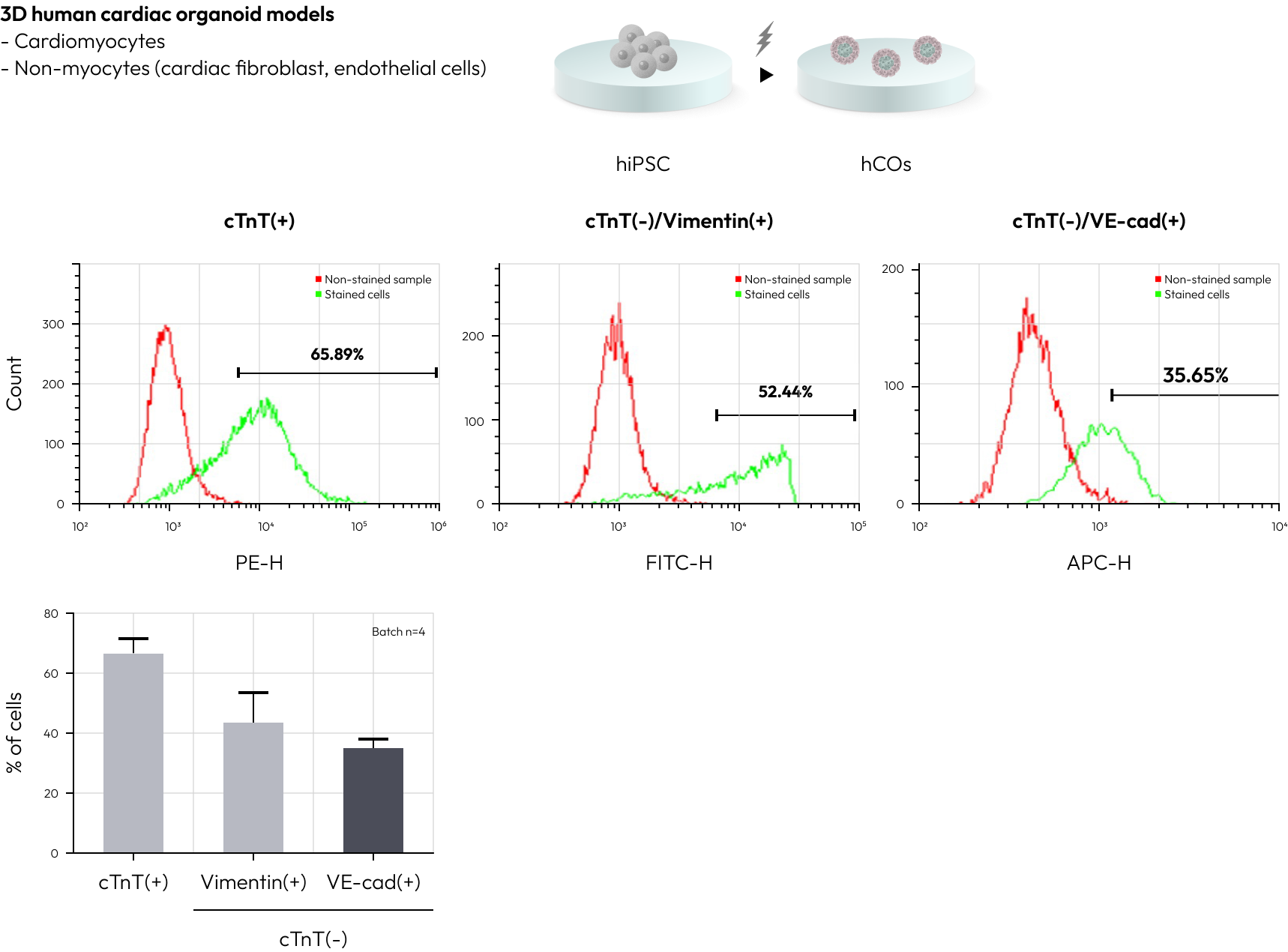
Characterization of cardiac organoid model
The cardiac organoids produced are composed similarly to the human heart, containing cardiomyocytes and non-muscle cells like cardiac fibroblasts and endothelial cells. Through FACS analysis, it was determined that cardiomyocytes constitute about 65% of the cell population, with the majority of the non-muscle cells being fibroblasts and endothelial cells.
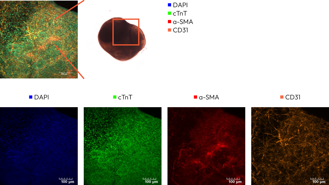
Using immunofluorescence, we have confirmed the expression of myocardial markers (cTnT, α-actinin), fibroblast markers (α-SMA), and endothelial cell markers (CD31) in our cardiac organoids. This method allows for detailed visualization and verification of specific cellular components critical to heart tissue function.
Patch clamp analysis
Our brain organoids can be observed in a structurally mature form and can be differentiated into the cerebrum and midbrain. Additionally, brain organoids are composed of various cells such as neurons and glial cells.
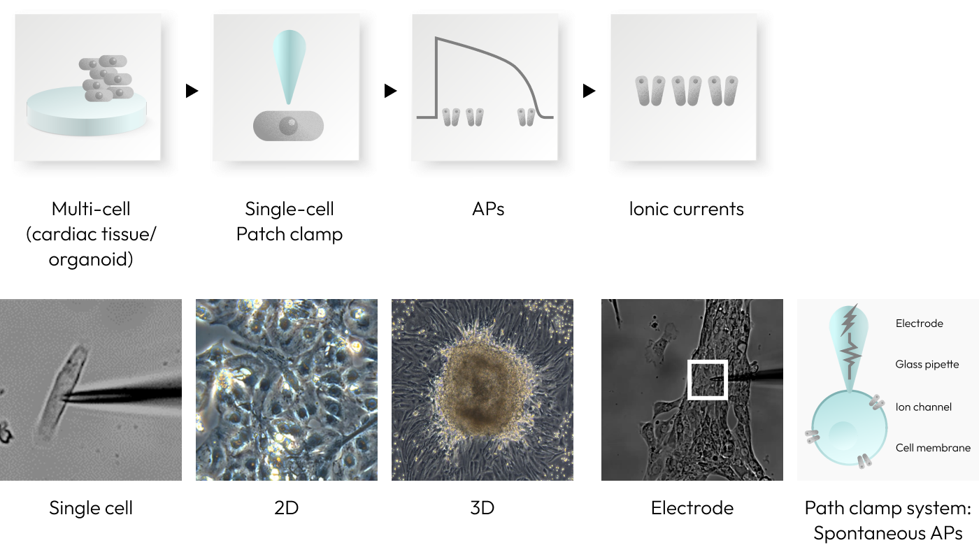
Real-time recording of action potentials
To analyzetheelectrophysiologicalproperties of cardiacorganoidswithspontaneousbeating, weemploythepatchclamptechnique. Byinsertingelectrodesintothecellmembranes of thecardiacorganoids, wecanmonitoractionpotentialsinreal-time, providing valuable insights into their functional behavior.
Cardiac organoids have electrophysiological properties similiar to cardiac tissue
Important Details
Validity: Until September 30th, 23:59 German time.
Applicability: One experiment per indication (Oncology, Skin & Cosmetics, Organoids).
Flexibility: Use the coupon unlimited times until the event ends.
Conditions: Applies when proceeding with a contract.
Log in to MyLab and
download your gift voucher from the ‘Vouchers’ menu!

")

